Back Bones Diagram / Pin By Sydney Physio Clinic On Relevant Anatomy Of Pelvis And Spine Human Spine Human Body Organs Thoracic Spine Mobility. Lower jaw (mandible) collar bone. It connects with the collarbone in the front of the body. The scapula, or shoulder blade , is a flat triangular bone located in the back of the shoulder. Click here to read about mesothelioma and its differential diagnosis and mesothelioma treatments. Money back guarantee refund in 15 days.
The temporal bone is one of the thickest bones in the skull. The bones of the chest — namely the rib cage and spine — protect vital organs from injury, and also provide structural support for the body. The outside of the flat bone consists of a layer of connective tissue called the periosteum. When a human finishes growing these parts fuse together. Diagram with back nerves and bones pain.
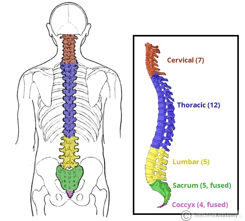
The Vertebral Column Joints Vertebrae Vertebral Structure from teachmeanatomy.info The vertebral column (also known as the backbone or the spine), is a column of approximately 33 small bones, called vertebrae. The human skeleton is a bony framework that not only gives shape to the body, but also. Diagram with back nerves and bones pain. It is the methodology used for. These bones have a marrow, but not a bone marrow cavity. We also discuss what are osteons, what are canaliculi, what are. The column runs from the cranium to the apex of the coccyx, on the posterior aspect of the body. It connects with the collarbone in the front of the body.
The column runs from the cranium to the apex of the coccyx, on the posterior aspect of the body.
Bone science human diagram anchor chart human body health back skeleton. In order to navigate out of this carousel please use your heading shortcut key to navigate to the next or previous heading. The vertebral column of the lower back includes the five lumbar vertebrae, the sacrum, and the coccyx. The temporal bone is one of the thickest bones in the skull. This shopping feature will continue to load items when the enter key is pressed. Development and function of the bones and joints: There also are bands of fibrous connective tissue—the ligaments and the tendons—in intimate relationship with the parts of the a diagram of the human skeleton showing bone and cartilage. The vertebral column (also known as the backbone or the spine), is a column of approximately 33 small bones, called vertebrae. In this video we discuss the structure of bone tissue and the components of bones. It is the methodology used for. Cheek bone (zygoma) upper jaw (maxilla). Disk herniation and gout, sciatica and spinal stenosis, osteoporosis illness set. The skeletal system includes all bones and joints of the human body.
These bones have a marrow, but not a bone marrow cavity. When a human finishes growing these parts fuse together. Disk herniation and gout, sciatica and spinal stenosis, osteoporosis illness set. The temporal bones are two major bones in the skull, or cranium. The skeletal system includes all bones and joints of the human body.
Back Muscles Anatomy Of Back Pain In Diagrams Goodpath from images.ctfassets.net 12 photos of the human back bones diagram. These bones work together to provide flexibility to the trunk, support the muscles of the trunk, and protect the spinal cord and spinal nerves of the back. Continue scrolling to read more below. Are you searching for bone diagram png images or vector? The vertebral column (also known as the backbone or the spine), is a column of approximately 33 small bones, called vertebrae. Master fuse box manitou wiring diagrams mazda fuse box marathon wiring schematics marine engine diagram mazda b2200 wiring maytag wiring diagram dryer map sensor wiring diagram. This framework consists of many individual bones and cartilages. We also discuss what are osteons, what are canaliculi, what are.
Are you searching for bone diagram png images or vector?
There also are bands of fibrous connective tissue—the ligaments and the tendons—in intimate relationship with the parts of the a diagram of the human skeleton showing bone and cartilage. A bone is a rigid tissue that constitutes part of the vertebrate skeleton in animals. Click here to read about mesothelioma and its differential diagnosis and mesothelioma treatments. Lower back of the head. Bones protect the various organs of the body, produce red and white blood cells, store minerals. Money back guarantee refund in 15 days. The scapula, or shoulder blade , is a flat triangular bone located in the back of the shoulder. Back anatomy diagram lower bones rear view of human skeletal system showing upper back stock photo anatomy of the spine and back anatomy of the back bones sciences. The lower back is also associated with feeling unsupported (not backed up) by a family member, partner, friend, teacher, colleague, or employer. Diagram with back nerves and bones pain. These bones have a marrow, but not a bone marrow cavity. Study this image showing the main bones of the body, then test your knowledge with our unlabeled diagram (download below). It is not possible to illustrate all of the bones and parts of bones that may be included on such courses without also including more complicated diagrams illustrating sections cut through the skull (such as back and base of the cranium, forms the back of the skull.
The human skeleton is a bony framework that not only gives shape to the body, but also. It connects with the collarbone in the front of the body. A bone is a rigid tissue that constitutes part of the vertebrate skeleton in animals. Choose from 14000+ bone diagram graphic resources and download in the form of png, eps, ai or psd. Back, bones and human spin diseases explanation vector.
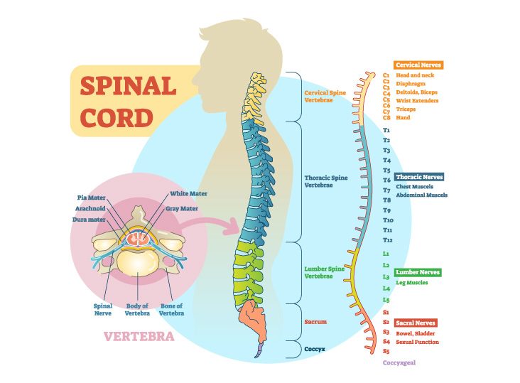
How The Spinal Cord Works Orthopedic Sports Medicine from orthosportsmed.com Bone science human diagram anchor chart human body health back skeleton. The temporal bone is one of the thickest bones in the skull. Study this image showing the main bones of the body, then test your knowledge with our unlabeled diagram (download below). Click here to read about mesothelioma and its differential diagnosis and mesothelioma treatments. The human skeleton is a bony framework that not only gives shape to the body, but also. These bones work together to provide flexibility to the trunk, support the muscles of the trunk, and protect the spinal cord and spinal nerves of the back. The vertebral column of the lower back includes the five lumbar vertebrae, the sacrum, and the coccyx. The end of the long bone is the epiphysis and the shaft is the diaphysis.
Diagram with back nerves and bones pain.
The human skeleton is a bony framework that not only gives shape to the body, but also. This framework consists of many individual bones and cartilages. These bones have a marrow, but not a bone marrow cavity. When a human finishes growing these parts fuse together. This shopping feature will continue to load items when the enter key is pressed. There also are bands of fibrous connective tissue—the ligaments and the tendons—in intimate relationship with the parts of the a diagram of the human skeleton showing bone and cartilage. We also discuss what are osteons, what are canaliculi, what are. Fishbone diagram or ishikawa diagram is a modern quality management tool that explains the cause and effect relationship for any quality issue that has arisen or that may arise. Bones protect the various organs of the body, produce red and white blood cells, store minerals. Fishbone diagram man method machine. Bone science human diagram anchor chart human body health back skeleton. Diagram with back nerves and bones pain. The vertebral column of the lower back includes the five lumbar vertebrae, the sacrum, and the coccyx.
Share :
Post a Comment
for "Back Bones Diagram / Pin By Sydney Physio Clinic On Relevant Anatomy Of Pelvis And Spine Human Spine Human Body Organs Thoracic Spine Mobility"


Post a Comment for "Back Bones Diagram / Pin By Sydney Physio Clinic On Relevant Anatomy Of Pelvis And Spine Human Spine Human Body Organs Thoracic Spine Mobility"